OCULAR PERFUSION
THE CAUSE OF Primary Open Angle Glaucoma
by Somkiat Athikhomkulchai
, M.D.
An Ophthalmologist
1. The Trabecular Meshwork
The
anterior part of the eyeball, there is a circulating, water-like fluid called
the aqueous humor. It is produced by the eye's ciliary body and initially excreted
into the posterior chamber and then flow through the pupil into the anterior chamber
, it leaves the eye mainly via the trabecular meshwork into the canal of Schlemm
which is connected to veins that course through the scleral surface.
Aqueous humor is actively
produced by the eye which the outflow has to overcome a certain amount of resistance.
This is the source of intraocular pressure, the IOP results from the balance between
the production and outflow of the aqueous humor.
In glaucoma patients,
the trabecular meshwork becomes increasingly laden with substances that impair
aqueous humor drainage. The increase in outflow resistance at trabecular meshwork
cause elevated IOP. The higher IOP induce the higher retinal venous pressure and
thus reduce retinal arterial perfusion that lead to ischemia and retinal ganglion
cell death. That is, High IOP is a factor that impede
Ocular Perfusion.
2. The Lamina Cribrosa
The posterior part
of the eyeball, there is a thin mesh-like elastic network rich in collagen, called
the Lamina cribosa. It is the continuation of the scleral layer of the eyeball
and has many tiny holes that traversed by optic nerve fibers and blood vessels.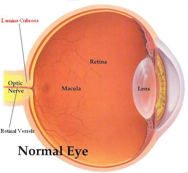
The physiological function
of the Lamina cribosa
2.1
To seal around optic nerve fibers, retinal artery and retinal vein, preventing
fluid leakage out of the eyeball, for the purpose to maintain the ocular tension.
2.2
To absorb pulsatile force of the retinal arterial blood, when reach the optic
nerve head before entering into the eyeball, by the elastic property of Lamina
cribosa around the artery. The pulsation of retinal artery, if existed, may interfere
the visual impulse sending to the brain.
2.3
To increase retinal venous pressure by the tightening force around the vein. The
pressure inside the retinal veins have to be at least as high as the IOP-or these
vessels would collapse. If the wrapping is too tight or getting tough, it cause
hemivein pulsation in someone.
Collagen changes in
Lamina cribosa whether from senile degeneration or genetic factor may impede axoplasmic
and vascular flow . This is another factor that
impede Ocular Perfusion.
In conclusion there are 2
factors that cooperate to be the cause of POAG
1.
the lamina cribosa and the impediment of blood supply
2.
The trabecular meshwork and the impediment of aqueous
outflow with increased ocular tension that impede blood supply
The
role of these 2 factors bring to the 3 types of POAG
1.The
high tension glaucoma
The
degenerative change of Lamina
cribrosa and Trabecular meshwork are the cause of this type
of glaucoma. Inadequate ocular perfusion leading to ganglion cells ischemia
and progressively die
Treatment
1.1
Stretch out the Lamina cribrosa plate to widen the pore that traversed by the
Retinal artery, enhancing ocular perfusion
1.2
Lowering the IOP to decrease the resistance of
ocular perfusion.
Nowadays
, all Glaucoma patients get only IOP lowering methods ( Medication, Laser or
Surgery ) without any treatment upon the Lamina cribrosa. This is the reason
to explain why some of these patients still go blind with all modalities of
IOP lowering method.
Eye
Massage can solve this problem because the procedure can
stretch out and widen the Lamina cribrosa pores together with decreasing the
IOP.
2.The
normal tension glaucoma
The
degenerative change of Lamina
cribrosa is the only and definite cause of this type of Glaucoma.
The IOP may be normal or low that is why most patients of this group do not
respond to all modalities of IOP lowering treament. Eye
Massage is the only and the definite
method to help these patients from going blind.
3.The
ocular hypertension
There
is degenerative change of Trabecular
meshwork and lead to elevated IOP but the ocular perfusion through
the Lamina cribrosa is good enough to supply adequate amount of blood for all
Ganglion cells. There is no Glaucomatous damage
and no need for any treatment, follow-up examination should
be done regularly to detect the Glaucomatous damage and when the treament is
reasonable.
EYE MASSAGE
This method can start
as Complementary treament without
any effect to the traditional method. After a month of treatment the patients
can realize whether to do Eye Massage
as the only treament or still couple with the traditional method.
-->Return
to Dr. Somkiat's Main Page<--